Medical SEM Style: Corneal Endothelial Cells
Details
Download Files (1)
Model description
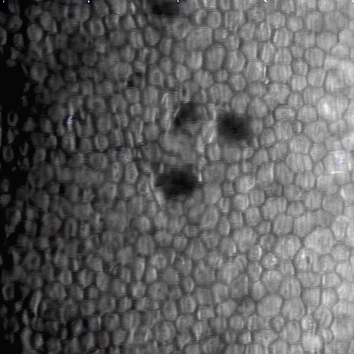
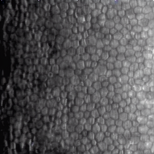
This LoRA generates photomicrographs of corneal endothelial cells in specular microscopy style (grayscale).
The model is trained to generate two distinct states:
Normal Cells: Regular hexagonal honeycomb pattern.
Cells with Guttata: Characteristic dark spots/drop-like lesions indicative of endothelial stress or dystrophy.
Recommended Weight: 1.0 Base Model: SD 1.5
Training Configuration:
Dataset: 1,000 corneal endothelial cell photomicrographs.
Resolution: All images resized to 512x512.
Training Strategy: 40 repeats per image.
Batch Size: 2